* Pustular skin lesions: The affected skin area invaded by larvae shows pustular lesions, containing numerous larvae. The two main causative agents are Dermatobia hominis and Cordylobia anthropophaga. During examination, there may be visible larvae, with a depressed central area, sometimes noticing small black breathing holes of the larvae or indirectly observing them through bubbles of secretions, which often consist of a mixture of blood and serum or pus. Patients often experience itching and sensation of something moving inside, with increased pain at night. Additionally, other skin lesions such as vesicles, abrasions, and ulcers may be present. With treatment, the lesions can heal completely, though some patients may be left with scars or increased skin pigmentation. However, these lesions provide favorable conditions for bacterial superinfection. Other organ symptoms are less common, but patients may experience swollen lymph nodes in the surrounding area, fever, and other symptoms commonly associated with secondary superinfection.
.jpg)
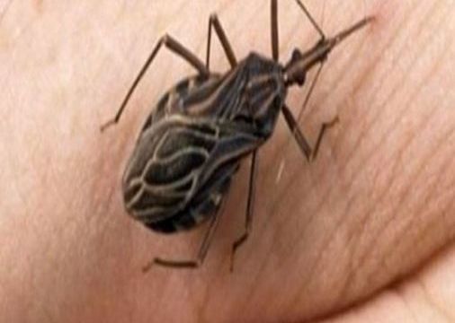
The skin areas invaded by larvae show pustular lesions.
* Injuries from larval movement: Initial injuries resemble pustular skin lesions; however, after a while, as larvae move, they invade the upper layers of the skin, creating red lesions, winding paths ranging from 1 cm to several tens of centimeters in length, raised edges above the skin surface, gradually fading in color towards the end of the path. Additionally, pustules, secretions, and secondary swelling may occur. Patients often feel itching, stinging sensations, and significant discomfort. If not diagnosed and treated promptly, larvae can migrate to other organs, burrowing deeper into the body.
* Disease at wound sites: Wounds often bleed, exude fluids, bleed and necrose. The wounds are often tortuous, sometimes forming tunnels, secreting blood, pus, emitting foul odors. Beneath the damaged and destroyed tissues, bacterial superinfection often occurs. Patients exhibit signs of infection such as fever, dirty tongue, foul breath, swollen nearby lymph nodes,…
– Nasal sinus and throat myiasis: Clinical symptoms often vary depending on the site of the lesion. In the mouth, symptoms such as pain, swelling, bad breath may occur; in some cases, larvae dying in the oral mucosa can cause painful swelling that may be misdiagnosed as a salivary gland tumor. If not diagnosed and treated promptly, this can lead to mouth ulcerations, bacterial superinfections, especially anaerobic bacteria. In the nose, irritation and increased nasal discharge are common, accompanied by nasal swelling, a sensation of foreign bodies in the nose, nosebleeds, or foul-smelling nasal discharge, often accompanied by fever. Myiasis in the ear causes patients to feel crawling sensations in the ear, often accompanied by frequent buzzing noises, ear bleeding, discharge from the ear, itching sensation, ringing in the ears, dizziness, hearing loss. The ear discharge is abundant, foul-smelling, and dirty. Larvae can penetrate deeper into brain tissue.

When myiasis affects the throat, patients often feel a sensation of foreign bodies in the throat, itching, and a burning sensation accompanied by coughing (often dry, sometimes productive), and a hoarse voice.
– Genitourinary myiasis:Patients may experience vaginal and vulvar inflammation with symptoms of foul-smelling discharge and itching, burning in the genital area. In males, inflammation of the foreskin, urethra, genital ulcers may occur,… Internal genital organs are less commonly affected than external genital organs.
– Gastrointestinal myiasis:The cause is ingestion of contaminated food or water containing larvae. Patients may experience abdominal pain, nausea, vomiting, diarrhea, anal itching, or bleeding in the lower digestive tract.
– Ocular myiasis:Patients experience eye pain, photophobia, decreased vision, eye swelling, tearing,… Examination may reveal conjunctival hemorrhage, bleeding, abundant pseudomembranes,…
– Central nervous system myiasis: Relatively rare but leaving sequelae and high mortality rates. Manifestations of brain and meningeal tissue damage, frontal lobe damage often accompanied by increased intracranial pressure and hydrocephalus.
Complications of Myiasis
Possible complications include: destruction and deformation of body tissues affecting organ function such as visual impairment, even blindness, decreased mobility, loss of skin integrity, brain tissue damage posing life-threatening risks,…; causing secondary superinfections such as bacterial or fungal infections; creating sinus tracts from infected organs;…
Transmission Route of Myiasis
The main transmission route is through ingestion. Humans can become infected by consuming food or water containing myiasis larvae. Botflies often concentrate in areas with poor sanitation or on animal bodies, and they can move around to deposit eggs, contaminating food and water. Additionally, direct contact with the wounds, sinus tracts of infected individuals can facilitate disease transmission.
At-Risk Population for Myiasis
As mentioned above, the main transmission route of the disease is through ingestion. Therefore, some at-risk populations are often those living in areas with poor living conditions and hygiene; those living and working in fly-infested environments, frequently consuming contaminated food and water, undercooked or unboiled; those with untreated or improperly cared for skin wounds or mucous membrane injuries; those in direct contact with the wounds or sinus tracts of myiasis-infected individuals; travelers to areas or regions with high prevalence of the disease.

Mucosal injuries not being properly cared for and maintained hygienically.